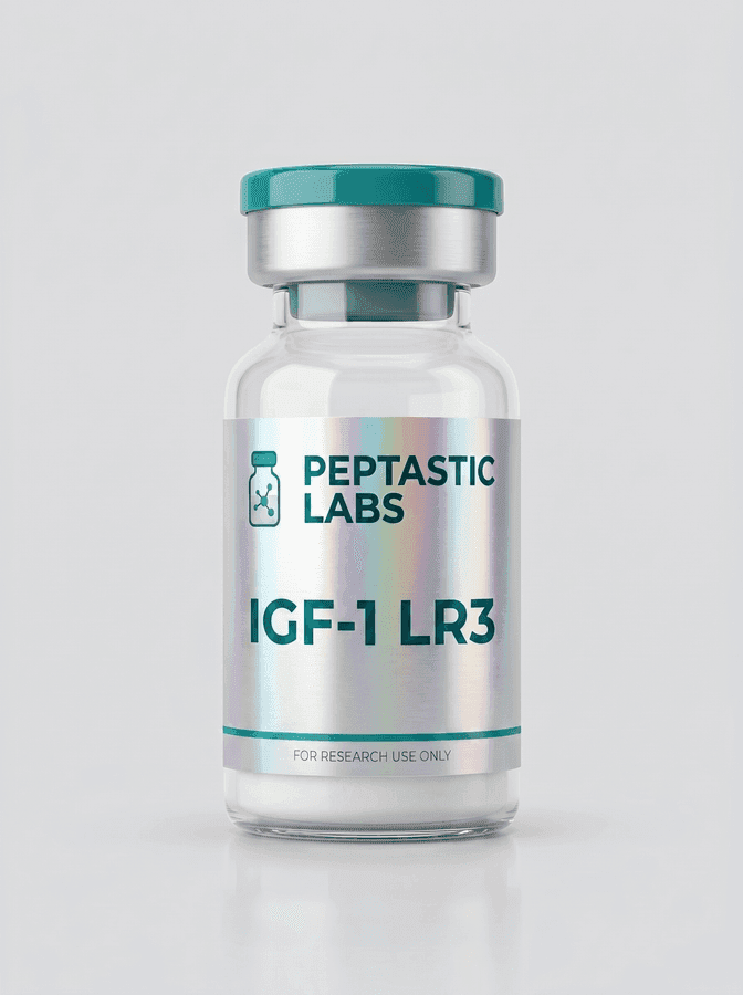
IGF-1 LR3 Cell Culture Research: Key Variables
· Peptastic Labs

A pronounced change in cell number, morphology or pathway readout after exposure to an IGF analogue can be compelling, but it is not automatically a clean measure of direct receptor activity. IGF-1 LR3 cell culture research is most useful when the material, model and assay conditions are documented well enough to separate a biological signal from serum effects, baseline pathway activation and routine culture variation.
IGF-1 LR3 is a long-acting insulin-like growth factor-1 analogue widely discussed in work involving cell growth, survival signalling, protein synthesis and metabolic regulation. Its altered structure can reduce interaction with some IGF-binding proteins relative to native IGF-1, potentially changing the free fraction available to cells in vitro. That property makes it scientifically interesting, but it also makes experimental context particularly consequential.
Why IGF-1 LR3 behaves differently in culture
Native IGF-1 activity is shaped by more than receptor binding. In biological fluids and many culture systems, IGF-binding proteins can sequester IGF ligands, influence ligand presentation and alter the duration of exposure. IGF-1 LR3 was developed with structural modifications that can lower binding to these proteins. In a cell culture setting, that may lead to a different apparent potency or time course than native IGF-1.
The key word is may. Outcomes depend on the cell type, its expression of IGF-1 receptor and insulin receptor isoforms, endogenous ligand production, medium composition and the presence of serum or protein supplements. A response observed in myoblasts, for example, should not be treated as a direct prediction of behaviour in fibroblasts, neuronal cells or tumour-derived lines.
IGF-1 receptor signalling commonly engages PI3K-AKT-mTOR and RAS-RAF-MEK-ERK-associated pathways. Those networks sit at the intersection of proliferation, survival, glucose handling, translation and differentiation. They are also influenced by insulin, epidermal growth factor, amino-acid availability, cell density and stress. For this reason, an increase in a downstream phosphoprotein alone does not establish that one pathway, or one receptor, explains the phenotype.
Building a defensible IGF-1 LR3 cell culture research plan
A sound study begins with a defined research question. Is the objective to assess an early signalling event, compare ligand sensitivity between cell models, examine proliferation over several days, or test whether a defined stressor changes IGF responsiveness? Each question calls for a different endpoint and time horizon.
Early pathway studies should distinguish transient receptor-proximal activity from later adaptive responses. Longer experiments need careful attention to nutrient depletion, confluence, medium changes and cumulative growth differences. When cells become crowded or metabolically stressed, signalling patterns can shift substantially even without experimental treatment.
A useful design typically compares vehicle control, native IGF-1 where relevant, and IGF-1 LR3 under the same culture conditions. Including a positive control appropriate to the pathway and a mechanism-informed inhibitor condition can further strengthen interpretation. Controls do not merely make a dataset look complete. They show whether the observed effect is ligand-specific, assay-specific or driven by broader culture conditions.
Concentration-response and time-course work is often more informative than selecting a single exposure condition. A single point can exaggerate a narrow effect or miss a delayed one. However, broader designs require enough replicates and a pre-specified analysis plan to avoid treating random variation as a biological trend.
Serum is not a background detail
Serum-containing media can introduce substantial variability into IGF-related experiments. Serum may contain endogenous growth factors, binding proteins and nutrients that influence baseline receptor activity and alter how a test ligand is presented to cells. Lot-to-lot variation can therefore affect both the magnitude and reproducibility of a result.
Serum reduction can improve interpretability in some models, but it is not universally preferable. Cells may experience stress, altered viability or differentiation changes under reduced-serum conditions. The appropriate choice depends on the biology being examined. Whatever condition is used, it should be defined clearly and applied consistently across comparison groups.
Cell identity and passage history matter
Misidentified or contaminated cell lines can invalidate otherwise careful work. Routine confirmation of cell identity, mycoplasma status, passage range and morphology is especially relevant in growth-factor research, where subtle changes in cell health can be mistaken for treatment effects.
Primary cells introduce another layer of variation. Donor source, isolation history and population heterogeneity may be central to the research question, but they should be treated as biological variables rather than hidden noise. A finding reproduced across independent donor preparations carries a different weight from one derived from a single culture.
Choosing meaningful readouts
The strongest studies pair a mechanistic readout with a functional one. For example, receptor or downstream phosphorylation can provide evidence of early pathway engagement, while cell counts, viability measures, differentiation markers or metabolic assays can show whether that engagement corresponds with a measurable phenotype.
No individual assay should carry the entire conclusion. Metabolic activity assays may reflect shifts in mitochondrial activity rather than cell number. Increased DNA synthesis does not necessarily mean sustained proliferation. Changes in protein abundance can be influenced by cell density and extraction quality. Orthogonal measurements reduce the chance of over-interpreting any one result.
Microscopy remains valuable here. Consistent imaging can reveal changes in confluence, cell shape, detachment or differentiation state that bulk assays may conceal. Images should support quantitative analysis, not replace it, and exposure settings should remain consistent between groups.
Material quality is part of the experimental variable
For peptide research, the stated identity of a vial is only the beginning. Purity, lot traceability, handling history and supporting analytical documentation can all influence confidence in an experiment. A Certificate of Analysis tied to the specific lot helps researchers assess what was supplied, while independent purity testing provides an additional quality-control reference.
It is also sensible to retain lot records in laboratory notes. If a result is unexpectedly strong, weak or difficult to reproduce, the ability to trace the material back to batch-specific documentation is practical scientific protection. A QR-linked or lot-matched record is more useful than generic paperwork that cannot be connected to the material used.
Peptide handling deserves the same discipline. Repeated freeze-thaw cycles, avoidable adsorption to unsuitable surfaces and inconsistent reconstitution practices can all complicate apparent activity. Laboratories should follow the supplier's storage guidance, use compatible laboratory consumables and record preparation details in a way another researcher can reproduce.
Peptastic Labs positions documented peptide materials as research use only, with batch-specific Certificates of Analysis and third-party testing intended to support traceable laboratory procurement. Such documentation does not substitute for good experimental design, but it gives researchers a clearer basis for evaluating one important source of variation.
Interpreting results without overstating them
IGF-1 LR3 has clear relevance to research on muscle cell biology, tissue repair pathways, cell survival and metabolic signalling. Yet in vitro findings are inherently bounded. A cultured-cell response does not establish effects in a whole organism, does not account for tissue distribution or systemic feedback, and does not provide a basis for human use.
The difference matters because IGF signalling is tightly regulated across tissues. Receptor abundance, local binding proteins, nutrient status, endocrine inputs and feedback mechanisms all shape activity outside a dish. Cell culture is best viewed as a controlled system for testing a defined hypothesis, not a shortcut around more complex biological questions.
Researchers also need to be cautious with language. “Associated with increased signalling under these conditions” is often more accurate than “causes growth” or “improves repair”. Precision makes results more useful to other laboratories and protects the distinction between preclinical research materials and products intended for therapeutic or personal use.
A well-documented negative result can be just as valuable as a positive one. If IGF-1 LR3 does not produce a measurable response in a validated model, that outcome may identify a meaningful boundary condition involving receptor expression, serum composition, differentiation state or assay timing. The most useful next step is rarely a bigger claim. It is a better-controlled question.
For research use only. Not for human or veterinary use. Not for consumption. Nothing in this article is medical advice or a recommendation for use in humans or animals.